
X-Ray - General Information
Imaging Services
X-Ray is the oldest diagnostic imaging procedure, and remains one of the most effective for its range of diagnostic applications. It uses small amounts of radiation that pass through the selected part of the body to produce an image. This procedure is often used to evaluate the chest, musculoskeletal system, and, if used with a contrast agent, the gastrointestinal system.
X-ray can evaluate abnormalities in the chest including lung cancer, pneumonia, fluid surrounding the heart, and pulmonary embolism; the gastrointestinal and urinary tracts for detection of everything from blockages and stones to ulcers and tumors; and the musculoskeletal system when bone or joint injuries or disease are suspected.
What to Expect
These tests typically take 15 to 30 minutes to complete. Usually, you will not need to do anything special to prepare for an X-ray exam. However, because of the technology involved, X-rays are hardly ever performed on a pregnant woman.
Follow any instructions given on the referral sheet you received from your physician. Like most other imaging exams, all metallic items including jewelry must be removed as they could interfere with the test.
Positioning of the body for the exam depends on the part of the body to be studied. Typically, you'll be lying down or standing up and must be very still while the images are taken. Several different views are obtained to see various angles.
If your X-ray exam includes using a contrast agent to examine the lower gastrointestinal or urinary tracts, prior preparation will be needed. Fasting and possibly using an enema or laxative the night before the exam may be required. You will be given complete instructions prior to your appointment.
We have imaging clinics in Placerville and Cameron Park. Please contact either location for more information.
Our Locations
-
 Diagnostic Imaging - Cameron Park 3581 Palmer DriveMap & Directions
Suite 300
Cameron Park, CA 95682 -
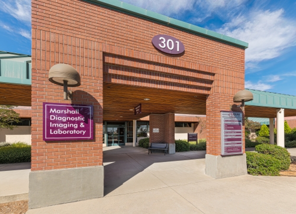
.1).2503130952078.jpg) Diagnostic Imaging - Placerville 1100 Marshall WayMap & Directions
Diagnostic Imaging - Placerville 1100 Marshall WayMap & Directions
Placerville, CA 95667 -
Marshall Hospital - Placerville 1100 Marshall WayMap & Directions
Placerville, CA 95667

.1).2503130952078.jpg)
See What Patients Are Saying About Marshall
-
“A huge thank you to all the staff – nurses, doctors and attendants – who took care of our dad while he was in the hospital for a week. Everyone we came in contact with was helpful, professional and ...”
-
Our family is so appreciative
“Our family is so appreciative of all the care and compassion we received during my breast cancer lumpectomy process. You all make healing so much easier and faster. Our family thanks you all.”- N.R. -
These folks make the process of going through cancer and all the meds quite easy.
“These folks make the process of going through cancer and all the meds quite easy. For me it was easy, and I could walk out and drive home after the treatments, I am thinking I'm lucky.”- C.M. -
I would recommend it for anyone who wants great medical care.
“Marshall is a really good medical center containing all necessary departments needed. The medical care given by my PCP is really top in class. In fact, all of the people I have come into contact with ...”- KB -
The whole staff is so nice and they truly want to help.
“I love the staff here. They always follow through with what they say they will do. I've never had to remind them. They are amazing. The whole staff is so nice and they truly want to help.”- AF -
I would like to thank the ER team for my amazing care I received.
“I would like to thank the ER team for my amazing care I received. I was treated with respect and caring throughout. The Dr. was very thorough with her work up. I finally got an accurate diagnosis for ...”- K -
“Marshall is filled with great doctors, and caring staff and nurses. It feels like family here.”- JH
-
“I sat in the ER waiting room for about 5 seconds before they were triaging me. The nicest most professional staff ever. I’m talking about everyone, not just the nurses and doctors. God bless all of ...”- Cory
-
“I had an MRI and I was very nervous. The radiologist was terrific! Kind, patient and got me through it, all calm and collected. Thank you!”- JL
